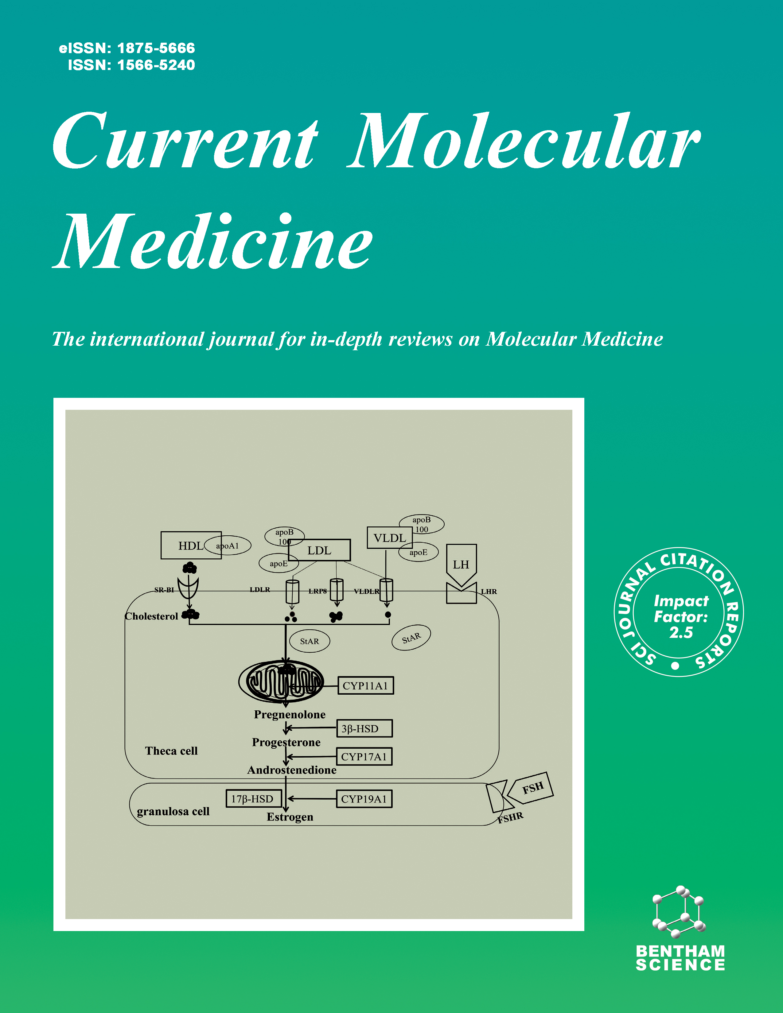
Current Molecular Medicine - Volume 17, Issue 1, 2017
Volume 17, Issue 1, 2017
-
-
The Immune Function of Ly6Chi Inflammatory Monocytes During Infection and Inflammation
More LessMonocytes originate from progenitors in bone marrow and circulate through blood vessels and patrol the vascular endothelium or differentiate into mononuclear phagocytes and infiltrate via the bloodstream to peripheral tissues during infection and other inflammatory conditions. Recruitment of inflammatory monocytes is essential for effectively clearance of bacteria, control of Toxoplasma gondii infections and limiting tissue damage. Unexpectedly, Ly6Chi inflammatory monocytes acquire the inflammatory regulatory capacity dual phenotypes during acute infection and inflammation. In addition, inflammatory monocytes can be recruited to tumor sites and inhibit tumor-specific immune defense. Here, we explored the recruitment and characteristics of Ly6Chi inflammatory monocytes and their immune regulatory function in acute inflammation, infection and tumor models. We also contrasted the differences between M2 macrophages and Ly6Chi inflammatory monocytes and, in particular, compared the shared common characteristics between these immune cell types. The Ly6Chi inflammatory monocyte regulatory functions in infectious and inflammatory conditions are the focus of this review.
-
-
-
Prion Function and Pathophysiology in Non-Mammalian Models
More LessAuthors: N. Guerrero, M. M. Meynard, J. Borgonovo, K. Palma, M. L. Concha and C. HetzMore than thirty years have passed since the discovery of the prion protein (PrP) and its causative role in transmissible spongiform encephalopathy. Since a combination of both gain- and loss-of-function mechanisms may underlay prion pathogenesis, understanding the physiological role of PrP may give important clues about disease mechanisms. Historically, the primary strategy for prion research has involved the use of human tissue, cell cultures and mammalian animal models. Nevertheless, experimental difficulties of in vivo studies and controversial observations obtained in these systems have stimulated the search for alternative animal models. PrPC is highly conserved in mammals, and PrPC-related orthologs are expressed in zebrafish, a vertebrate model organism suitable to study the mechanisms associated with human diseases. Invertebrate models, as they do not express PrPC have served to investigate the neurotoxic mechanisms of mammalian PrP. Here we overview most recent advances in the study of PrP function in normal and pathogenic conditions based on non-mammalian studies, highlighting the contribution of zebrafish, fly and worms to our current understanding of PrP biology.
-
-
-
Survival and Immunosuppression Induced by Hepatocyte Growth Factor in Chronic Lymphocytic Leukemia
More LessAuthors: P. Giannoni, G. Cutrona and D. de ToteroChronic lymphocytic leukemia (CLL), the most common leukemia among adults in the western world, is characterized by a progressive accumulation of relatively mature CD5+ B cells in peripheral blood, lymph nodes and bone marrow. Despite much recent advancement in therapy, CLL is still incurable. Lymph nodes and bone marrow represent sanctuary sites preserving leukemic cells from spontaneous or drug-induced apoptosis, and infiltration of leukemic cells in these districts correlates with clinical stages and prognosis. The central role played by the microenvironment in the disease has become increasingly clear. Different chemokines (CXCL12, CXCL13, CCL19, CCL21) may in fact participate in attracting CLL cells into bone marrow and lymph nodes, where various factors, such as IL-15 and CXCL12, enhance leukemic cells survival. Recently, we have suggested that hepatocyte growth factor (HGF), produced by microenvironmental stromal cells, can contribute to CLL pathogenesis. We have demonstrated that HGF exerts a double effect on CLL B cells through the interaction with its receptor c- MET; HGF, infact, protects CLL B cells, which are c-MET+, from apoptosis, and also polarizes mono/macrophages towards the M2 phenotype, thus facilitating the evasion of the CLL clone from immune control. This double effect appears mediated by the activation of two major signaling pathways: STAT3TYR705 and AKT. The aim of this review is to summarize data on HGF and c-MET expression in normal B cells and in B cell malignancies, with a particular emphasis on our results obtained in CLL. Altogether, the observations described here suggest that the HGF/c-MET axis may have a prominent role in malignancy progression further indicating novel potential therapeutic options aimed to block HGF-induced signaling pathways in B lymphoproliferative disorders.
-
-
-
Molecular Determinants of Colon Cancer Susceptibility in the East and West
More LessAuthors: W. M. Abdel-Rahman, M. E. Faris and P. PeltomakiThe currently available knowledge of factors that dictate the development and progression as well as the clinical outcome of colorectal cancers (CRC) is mainly derived from Western countries. Considerable number of publications document different incidence rates and contrasting clinical features of CRC in various groups such as the differences between urban vs. rural areas, young vs. old age and the East vs. the West. In particular, Egyptian CRC is a surprisingly young age disease with higher proportion of poorly differentiated and advanced stage cancers as compared to the Western counterparts. Less number of publications addressed the molecular genetics and epigenetic basis of these differences. The available data on CRC and other cancers support a substantial role of several environmental risk factors which impinge on the epigenome and alter the overall cellular and tissue homeostasis. Thus, environmental factors could play a role in predisposition to CRC in general as well as in shaping distinct disease phenotypes in different settings. On the other hand, the environment offers a wide range of preventive modalities including a selection of dietary chemopreventive agents which could play a significant role in fighting cancer at early stages. We here compare the clinical and molecular characteristics of Eastern and Western CRC based on the latest literature. The genetic, epigenetic and environmental etiologies for the observed differences are discussed. Finally, prospects for cancer prevention in light of the increased etiologic understanding are outlined.
-
-
-
Diabetes Type 4: A Paradigm Shift in the Understanding of Glaucoma, the Brain Specific Diabetes and the Candidature of Insulin as a Therapeutic Agent
More LessAuthors: M. A. Faiq and T. DadaIn the present analysis, we aim at probing into many important mechanisms that serve to bridge conceptual gaps to fill up the mosaic of a picture revealing that glaucoma indeed is brain specific diabetes and more appropriately "Diabetes Type 4". Based on this conceptual substance, we weave a novel idea of insulin being a potential remedy for glaucoma. This analysis synthesizes upon the published literature on brain changes in glaucoma, possibility of isolated brain diabetes, insulin signaling glitches in glaucoma pathology, mitochondrial dysfunction and insulin resistance in glaucomatous eyes, insulin mediated regulation of intraocular pressure and its dysregulation in mitochondrial dysfunction. We also look into the role of amyloidopathy and taupathy in glaucoma pathogenesis vis-a-vis insulin signaling. At every step, the discussion reveals that insulin and other allied moieties are a sure promise for glaucoma treatment and management. In this article, we aim at synthesizing a persuasive and all inclusive picture of glaucoma etiopathomechanism centered on "insulin-hypofunctionality" in the central nervous system (i.e. brain specific diabetes). We start with considering the possibility of neurodegenerative diabetes that exists independent of the peripheral diabetes. Once that condition is met, then a metabolic conglomeration of this brain specific diabetes is deliberated upon leading us to understand the development of retinal ganglion cell apoptosis, intraocular pressure elevation, optic cupping and mitochondrial dysfunction. All these are the hallmarks and sufficient conditions to satisfy the diagnostic criteria for glaucoma. Immediate application of this analysis points towards glaucoma therapy centered upon improving what we have termed insulin-hypofunctionality.
-
-
-
Molecular Determinants for STIM1 Activation During Store- Operated Ca2+ Entry
More LessBackground: STIM/ORAI-mediated store-operated Ca2+ entry (SOCE) mediates a myriad of Ca2+-dependent cellular activities in mammals. Genetic defects in STIM1/ORAI1 lead to devastating severe combined immunodeficiency; whereas gain-offunction mutations in STIM1/ORAI1 are intimately associated with tubular aggregate myopathy. At molecular level, a decrease in the Ca2+ concentrations within the lumen of endoplasmic reticulum (ER) initiates multimerization of the STIM1 luminal domain to switch on the STIM1 cytoplasmic domain to engage and gate ORAI channels, thereby leading to the ultimate Ca2+ influx from the extracellular space into the cytosol. Despite tremendous progress made in dissecting functional STIM1-ORAI1 coupling, the activation mechanism of SOCE remains to be fully characterized. Objective and Methods: Building upon a robust fluorescence resonance energy transfer assay designed to monitor STIM1 intramolecular autoinhibition, we aimed to systematically dissect the molecular determinants required for the activation and oligomerization of STIM1. Results: Here we showed that truncation of the STIM1 luminal domain predisposes STIM1 to adopt a more active conformation. Replacement of the single transmembrane (TM) domain of STIM1 by a more rigid dimerized TM domain of glycophorin A abolished STIM1 activation. But this adverse effect could be partially reversed by disrupting the TM dimerization interface. Moreover, our study revealed regions that are important for the optimal assembly of hetero-oligomers composed of full-length STIM1 with its minimal STIM1-ORAI activating region, SOAR. Conclusions: Our study clarifies the roles of major STIM1 functional domains in maintaining a quiescent configuration of STIM1 to prevent preactivation of SOCE.
-
-
-
Decreased HoxD10 Expression Promotes a Proliferative and Aggressive Phenotype in Prostate Cancer
More LessAuthors: R.-J. Mo, J-M Lu, Y.-P. Wan, W. Hua, Y.-X. Liang, Y.-J. Zhuo, Q.-W. Kuang, Y.-L. Liu, H.-C. He and W.-D. ZhongHoxD10 gene plays a critical role in cell proliferation in the process of tumor development. However, the protein expression level and the function of HoxD10 in prostate cancer remain unknown. Using tissue microarray, we demonstrate that the protein expression of HoxD10 is commonly decreased in prostate cancer tissues (n = 92) compared to adjacent benign prostate tissues (n = 77). Functionally, knockdown of HoxD10 resulted in significant promotion of prostate cancer cell proliferation. Moreover, knockdown of HoxD10 strikingly stimulated prostate tumor growth in a mouse xenograft model. We also found a significant association between decreased immunohistochemical staining of HoxD10 expression and higher Gleason score (P = 0.031) and advanced clinical pathological stage (P = 0.011). An analysis of the Taylor database revealed that decreased HoxD10 expression predicted worse biochemical recurrence (BCR)-free survival of PCa patients (P = 0.005) and the multivariate analyses further supported that HoxD10 might be an independent predictor for BCR-free survival (P = 0.027). Collectively, our data suggest that the loss of HoxD10 function is common and may thus result in a progressive phenotype in PCa. HoxD10 may function as a biomarker that differentiates patients with BCR disease from the ones that are not after radical prostatectomy, implicating its potential as a therapeutic target.
-
-
-
Evidence of PKC Binding and Translocation to Explain the Anticancer Mechanism of Chlorogenic Acid in Breast Cancer Cells
More LessAuthors: S. J. Deka, S. Gorai, D. Manna and V. TrivediChlorogenic acid (CGA) exhibits potentials towards liver, breast and skin cancer. Cancer cells stimulated with CGA exhibits differential expression of transcriptional factors and regulatory molecules but the molecular target of the molecule is not known. Superposition of biophoric elements of CGA with Curcumin gives maximum common substructure score of 0.90. Molecular modeling studies further suggest that CGA fits into the C1b domain of PKC with extensive interaction with residues lining binding site. It binds PKC in a concentration dependent manner with dissociation constant KD, 28.84±3.95 μM. PKC-CGA complex is stable with minimal distortion to the 3-D structure and maintains the hydrogen bonding between ligand and receptor during simulation period. Cells stimulated with CGA causes 12.1 ± 0.56% PKC translocation from the cytosol to the plasma membrane. It disturbs the cell cycle and arrest the cancer cell at the G1 phase with a reduction in S-phase. Chlorogenic acid exhibits killing of cancer cells in a dose-dependent manner with an IC50 of 75.88 ± 4.54μg/ml and 52.5 ± 4.72μg/ml towards MDAMB-231 and MCF-7 cells respectively. It induces apoptosis in cancer cells as evident by AO/EtBr staining and degradation of genomic DNA to give a laddering pattern. Apoptosis in cancer cells involves mitochondrial pathway as supported by a reduction in mitochondrial potentials and release of cyt-C into the cytosol. Hence, the current study has established PKC as an important signaling molecule to the observed anti-cancer effects of CGA and provides the impetus to design better CGA analogs for improved anti-cancer potential against the malignant tumor.
-
Volumes & issues
-
Volume 25 (2025)
-
Volume 24 (2024)
-
Volume 23 (2023)
-
Volume 22 (2022)
-
Volume 21 (2021)
-
Volume 20 (2020)
-
Volume 19 (2019)
-
Volume 18 (2018)
-
Volume 17 (2017)
-
Volume 16 (2016)
-
Volume 15 (2015)
-
Volume 14 (2014)
-
Volume 13 (2013)
-
Volume 12 (2012)
-
Volume 11 (2011)
-
Volume 10 (2010)
-
Volume 9 (2009)
-
Volume 8 (2008)
-
Volume 7 (2007)
-
Volume 6 (2006)
-
Volume 5 (2005)
-
Volume 4 (2004)
-
Volume 3 (2003)
-
Volume 2 (2002)
-
Volume 1 (2001)
Most Read This Month


