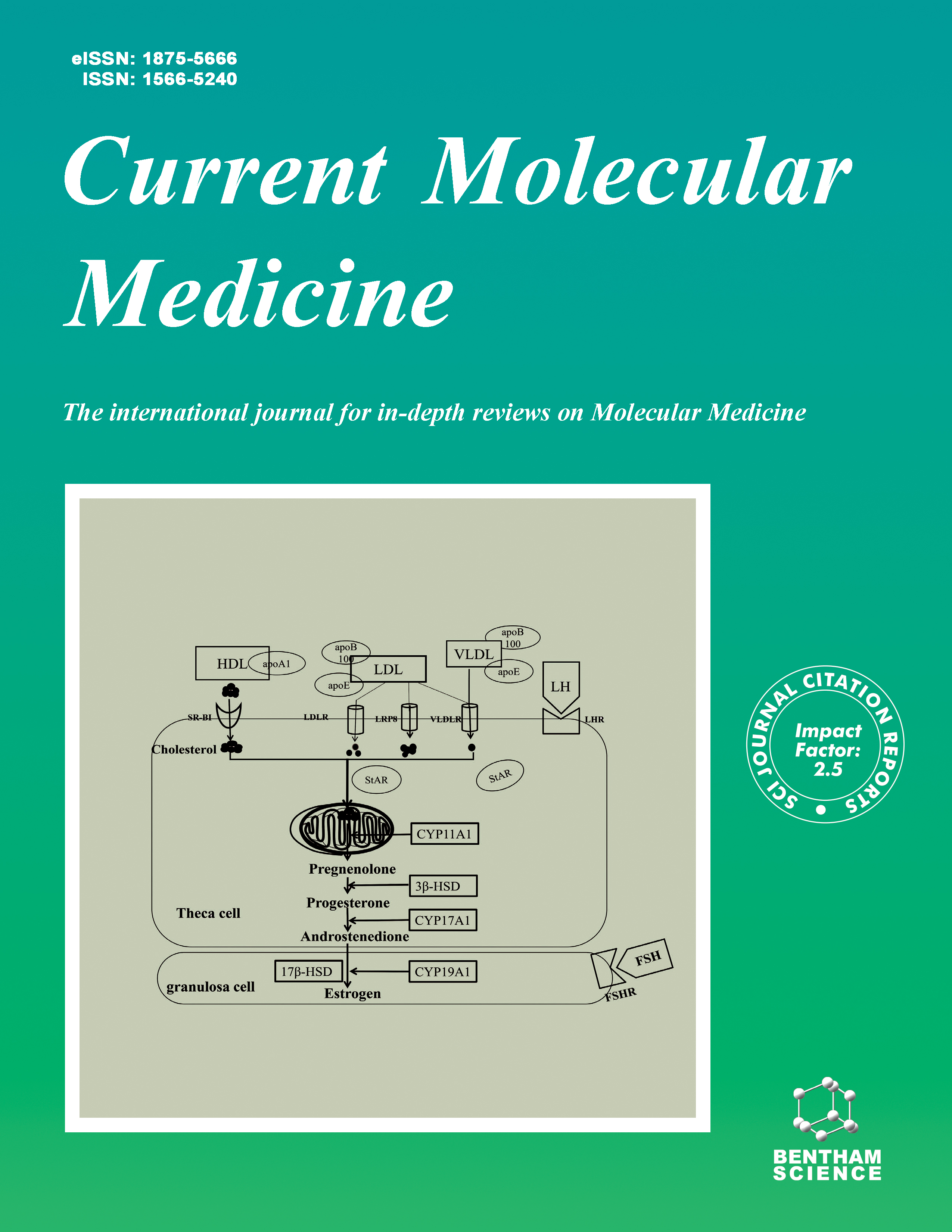
Current Molecular Medicine - Volume 15, Issue 9, 2015
Volume 15, Issue 9, 2015
-
-
Entosis and Related Forms of Cell Death within Cells.
More LessAuthors: Y. Wang and X.-D. WangBy eliminating the unneeded or mutant cells, programmed cell death actively participates in a wide range of biological processes from embryonic development to homeostasis maintenance in adult. Continuing efforts have identified multiple cell death pathways, with apoptosis, necrosis and autophage the mostly studied. Recently a unique cell death pathway called “cell-in-cell death” has been defined. Unlike traditional cell death pathways, cell-in-cell death, characterized by cell death within another cell, is triggered by the invasion of one cell into its neighbor and executed by either lysosome-dependent degradation or caspase-dependent apoptosis. With remarkable progresses on cell-in-cell over past few years, multiple mechanisms, including entosis, cannibalism and emperitosis, are found to be responsible for cell-in-cell death. Some key questions, such as specific biochemical markers to distinguish precisely the properties of different cell-in-cell structures and the physiological and pathological relevance, remain to be addressed. In light of this situation and a surge of interests, leading scientists in this field intend to share with readers current research progresses on cell-in-cell structures from different model systems through this special edition on cell-in-cell. The mechanistic advances will be highlighted while the future researches be speculated.
-
-
-
Cell-in-cell phenomenon: A New Paradigm in Life Sciences.
More LessBy X. WangCell-in-cell, a phenomenon characterized by one or more viable cells entering actively into another cell, was observed more than a century and has only attracted more attention in recent years and is becoming a new hot topic in the biological field, owing its biological significance in evolutionary as well as physiological and pathological relevance in development, homeostasis and diseases. In this paper we focus on the diversity, evolutionary conservatism and clinical implication of cell-in-cell as well as latest opinions on the research strategies. Based on the findings from our laboratory and other research groups three working models of cell-in-cell are also proposed.
-
-
-
Suicidal emperipolesis: a process leading to cell-in-cell structures, T cell clearance and immune homeostasis.
More LessAuthors: F. Sierro, S. S. Tay, A. Warren, D. G. Le Couteur, G. W. McCaughan, D. G. Bowen and P. Bertolino“Suicidal emperipolesis” is one of the most recently reported processes leading to cell-in-cell structures that promote cell death. This process was discovered in studies investigating the fate of autoreactive CD8 T cells activated within the liver. Recently, we reported that activated T cells invaded hepatocytes, formed transient cell-in-cell structures, and were rapidly degraded within endosomal/lysosomal compartments by a non-apoptotic pathway. Importantly, pharmacological inhibition of this process caused intrahepatic accumulation of tissue-reactive T cells and breach of immune tolerance. The characterization of the molecular mechanisms of suicidal emperipolesis is still in its infancy, but initial studies suggest this phenomenon is distinct from other reported cell-in-cell structures. As opposed to the formation of other cell-in-cell structures, suicidal emperipolesis takes place in a non-malignant environment, and without obvious pathology. It is therefore the first cell-in-cell structure described to have a role in maintaining homeostasis in normal physiology in higher organisms. T cell emperipolesis within hepatocytes has also been observed by pathologists in a range of chronic human liver pathologies. As T cell-in-hepatocyte structures resulting from suicidal emperipolesis are very transiently observed in normal physiology, their accumulation during liver disease would suggest that severe tissue injury is promoted by, or associated with, defective T cell clearance. In this review, we compare “suicidal emperipolesis” to other processes leading to cell-in-cell structures, and consider its potential biological roles in maintaining immune homeostasis and tolerance in the context of the hepatic environment.
-
-
-
Thymic Nurse Cells Participate in Heterotypic Internalization and Repertoire Selection of Immature Thymocytes; Their Removal from the Thymus of Autoimmune Animals May be Important to Disease Etiology
More LessAuthors: J. C. Guyden, M. Martinez, R. V.E. Chilukuri, V. Reid, F. Kelly and M.-O.D. SammsThymic nurse cells (TNCs) are specialized epithelial cells that reside in the thymic cortex. The initial report of their discovery in 1980 showed TNCs to contain up to 200 thymocytes within specialized vacuoles in their cytoplasm. Much has been reported since that time to determine the function of this heterotypic internalization event that exists between TNCs and developing thymocytes. In this review, we discuss the literature reported that describes the internalization event and the role TNCs play during T cell development in the thymus as well as why these multicellular complexes may be important in inhibiting the development of autoimmune diseases.
-
-
-
Cancer Cell Cannibalism: A Primeval Option to Survive.
More LessAuthors: F. Lozupone and S. FaisCancer cell cannibalism is currently defined as a phenomenon in which an ensemble of a larger cell containing a smaller one, often in a big cytoplasmic vacuole, is detected in either cultured tumor cells or a tumor sample. After almost one century of considering this phenomenon as a sort of neglected curiosity, some recent studies have first proposed tumor cell cannibalism as a sort of “aberrant phagocytosis”, making malignant cells very similar to professional phagocytes. Later, further research has shown that, differently to macrophages, exclusively ingesting exogenous material, apoptotic bodies, or cell debris, tumor cells are able to engulf other cells, including lymphocytes and erythrocytes, either dead or alive, with the main purpose to feed on them. This phenomenon has been associated to the malignancy of tumors, mostly exclusive of metastatic cells, and often associated to poor prognosis. The cannibalistic behavior increased depending on the microenvironmental condition of tumor cells, such as low nutrient supply or low pH, suggesting its key survival option for malignant cancers. However, the evidence that malignant cells may cannibalize tumor-infiltrating lymphocytes that act as their killers, suggests that tumor cell cannibalism could be a very direct and efficient way to neutralize immune response, as well. Tumor cell cannibalism may represent a sign of regression to a simpler, ancestral or primeval life style, similar to that of unicellular microorganisms, such as amoebas, where the goal is to survive and propagate in an overcrowded and very hostile microenvironment. In fact, we discovered that metastatic melanoma cells share with amoebas a transmembrane protein TM9SF4, indeed related to the cannibal behavior of these cells. This review attempts to provide a comprehensive description of the current knowledge about the role of TM9SF4 in cancer, highlighting its role as a key player in the cannibal behavior of malignant cancer cells. Moreover, we discuss differences and similarities between tumor cannibalism, entosis, phagocytosis and emperipolesis.
-
-
-
Phagoptosis - Cell Death By Phagocytosis - Plays Central Roles in Physiology, Host Defense and Pathology
More LessAuthors: G. C. Brown, A. Vilalta and M. FrickerCell death by phagocytosis – termed ‘phagoptosis’ for short – is a form of cell death caused by the cell being phagocytosed i.e. recognised, engulfed and digested by another cell. Phagocytes eat cells that: i) expose ‘eat-me’ signals, ii) lose ‘don’t-eat-me’ signals, and/or iii) bind opsonins. Live cells may express such signals as a result of cell stress, damage, activation or senescence, which can result in phagoptosis. Phagoptosis may be the most abundant form of cell death physiologically as it mediates erythrocyte turnover. It also regulates: reproduction by phagocytosis of sperm, development by removal stem cells and excess cells, and immunity by removal of activated neutrophils and T cells. Phagoptosis mediates the recognition of non-self and host defence against pathogens and cancer cells. However, in inflammatory conditions, excessive phagoptosis may kill our cells, leading to conditions such as hemophagy and neuronal loss.
-
-
-
Mammalian Cell Competitions, Cell-in-Cell Phenomena and Their Biomedical Implications.
More LessCell competition was first identified four decades ago as a mechanism to eliminate less fit cells during development in Drosophila melanogaster, and later postulated to be involved in tumorigenesis of human beings. However, evidence for a similar mechanism functional in mammals and tumors was missed until recent years. Like cell competition in fly, multiple forms of competition mechanisms were reported in mammalian system, and some of them were found participating in tumor initiation. Lately, entosis, a mechanism of cell cannibalisms responsible for the formation of cell-in-cell structures in human tumors, was identified as a novel member of ever-expanding family of mammalian cell competitions (MaCCs), and proposed to be able to promote clonal selection and tumor evolution. Thus, engulfment by neighboring cells other than the professional phagocytes, an issue still in debate in fly, was clearly demonstrated in mammals to be responsible for loser elimination. Competition mediated by cell-in-cell structures, formed by multiple cannibal mechanisms, constitutes a novel class of MaCCs. This review will summarize current research on mammalian cell competitions, followed by feature and mechanism analysis and their potential implications in the pathology and treatment of human tumors.
-
-
-
Entosis: Cell-in-Cell Formation that Kills Through Entotic Cell Death
More LessAuthors: O. Florey, S. E. Kim and M. OverholtzerEntosis is a cell-in-cell formation mechanism that targets viable cells for uptake in epithelial cell cultures and human tumors. Entotic cells control their own engulfment, by invading into their hosts in a Rho-GTPase and actomyosin-dependent manner. Although entotic cells are internalized while alive, most eventually undergo a non-apoptotic form of cell death, called entotic cell death, that is executed non-cell-autonomously by autophagy proteins and lysosomes. Here we review the current understanding of entosis and entotic cell death and discuss the potential roles of this process in cancer.
-
-
-
The physics for the formation of cell-in-cell structures.
More LessThe formations of cell-in-cell structures have been found in several important biological processes. Recent studies have shed light on the biochemical signaling pathways as well as the quantitative understandings of the underlying physics. Multiple new features that regulate the cellular engulfment have been identified. However, the driving forces promoting the structural formation are still under debate. This review focuses on the recent progress and discusses the potential significance of the existing physical models.
-
-
-
Emperipolesis is a potential histological hallmark associated with chronic hepatitis B.
More LessAlthough emperipolesis exists in infectious liver diseases, the diagnostic value of emperipolesis in chronic hepatitis B is not exactly known. The aim of this study is to evaluate the histological characteristics and laboratory parameters of emperipolesis in chronic hepatitis B. Totally 402 patients with hepatitis B and other liver diseases were processed in a retrospective assessment. Inflammatory severity of hepatitis B was evaluated with Ishak Scoring System. Immunofluorescent staining was performed for CD8 (T cells), CD20 (B cells), CD56 (NK cells), CD68 (macrophages) and MPO (neutrophils). Emperipolesis was observed in 74.0% of patients with chronic hepatitis B (CHB) and 82.8% of patients with acute hepatitis B (AHB). In emperipolesis, CD8+ T cell was the main cell type. Patients with emperipolesis in CHB got high scores of inflammatory activity. Among patients with CHB, emperipolesis was present with higher serum ALT, AST and GGT levels. HBV DNA Load in patients with emperipolesis was as 10 times high as those without emperipolesis. HBeAb was significantly correlated with the evidence of emperipolesis. In chronic hepatitis B, emperipolesis was associated with severity of liver injury. The presence of emperipolesis was an indicator of active liver inflammation.
-
Volumes & issues
-
Volume 25 (2025)
-
Volume 24 (2024)
-
Volume 23 (2023)
-
Volume 22 (2022)
-
Volume 21 (2021)
-
Volume 20 (2020)
-
Volume 19 (2019)
-
Volume 18 (2018)
-
Volume 17 (2017)
-
Volume 16 (2016)
-
Volume 15 (2015)
-
Volume 14 (2014)
-
Volume 13 (2013)
-
Volume 12 (2012)
-
Volume 11 (2011)
-
Volume 10 (2010)
-
Volume 9 (2009)
-
Volume 8 (2008)
-
Volume 7 (2007)
-
Volume 6 (2006)
-
Volume 5 (2005)
-
Volume 4 (2004)
-
Volume 3 (2003)
-
Volume 2 (2002)
-
Volume 1 (2001)
Most Read This Month


